
A lithium dendrite does not appear overnight. By the time a cell shorts, the deposit has usually been growing quietly on the anode surface for hundreds of cycles — leaving traces at every one of them, almost none of which show up on a capacity plot. Once it pierces the separator, all that is left to do is failure analysis.
This article walks through the life of a dendrite in order: where it nucleates, why it accelerates, how it converts cyclable lithium into dead lithium, and at which stage it can still be caught.
It starts with inhomogeneous deposition
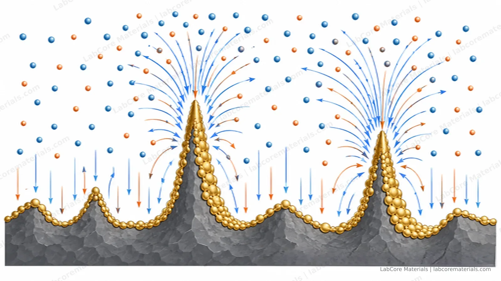
Non-uniform deposition is where the story begins. No anode surface is ideal: microscopic roughness, uneven SEI thickness and local differences in ion concentration make lithium deposit at different rates across the surface. Ions take the path of least resistance, and any protrusion concentrates the electric field onto itself — it deposits faster, grows taller, and attracts more ions. A positive feedback loop is born.
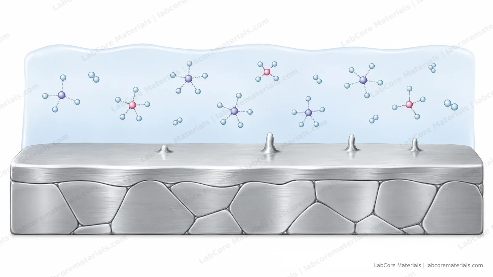
At this stage the dendrite is a microscopic bump. Intercalation anodes (graphite, silicon) face the same problem whenever their operating potential dips below lithium's deposition potential: once the local overpotential crosses the threshold, metallic lithium starts plating outside the active material. Fast-charge protocols are strict about temperature and current for exactly one reason — to postpone this starting point.
When growth accelerates
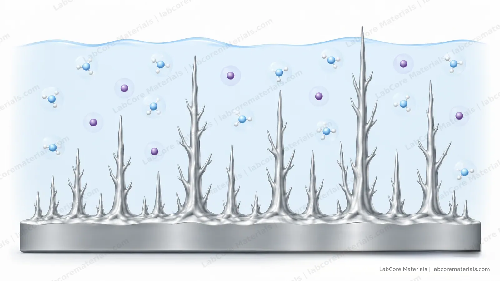
What governs the growth rate is the local lithium supply at the surface. High-rate charging depletes surface concentration and steepens the gradient, shifting deposition from uniform plating to localized extrusion; at low temperature, both interfacial kinetics and diffusion slow down, amplifying the inhomogeneity. Morphology follows the conditions: under mild ones the deposit is a soft, mossy layer; under harsh ones it grows a defined trunk — a dendrite in the strict sense, and a far greater threat to the separator.
Fracture and regrowth: the dead-lithium assembly line
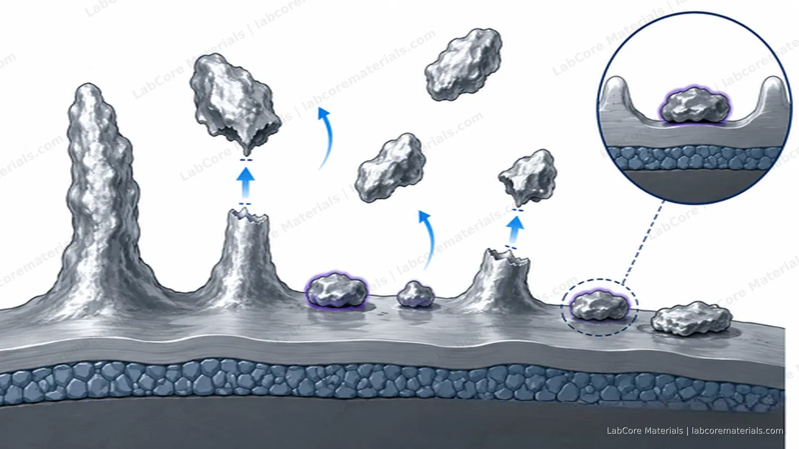
A dendrite is dressed in SEI. The faster it grows, the more often the SEI ruptures, and every rupture exposes fresh surface whose repair consumes fresh lithium and electrolyte. Stripping is worse: SEI-wrapped dendrites tend to break off at the root and lose electrical contact — physically still in the electrode, electrochemically gone. That is dead lithium.
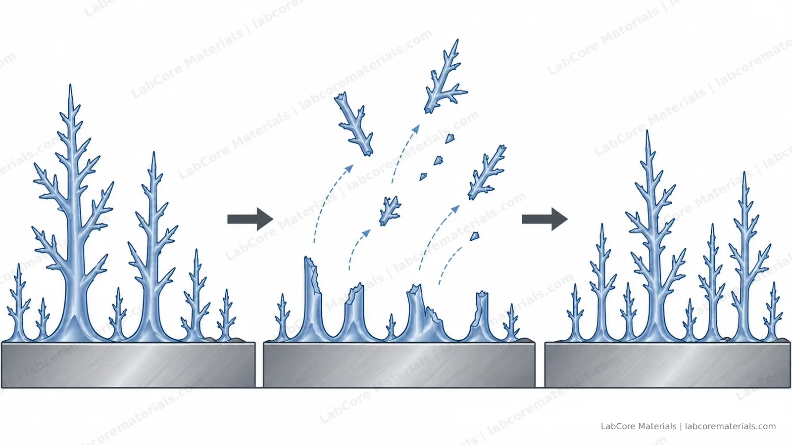
Every cycle repeats the sequence: grow, break, isolate. Under severe plating, a substantial share of the capacity loss is not "unused" lithium but lithium manufactured into dead lithium — which is why plating-induced fade is nearly irreversible, and a rest does not bring it back.
The endpoint: piercing the separator
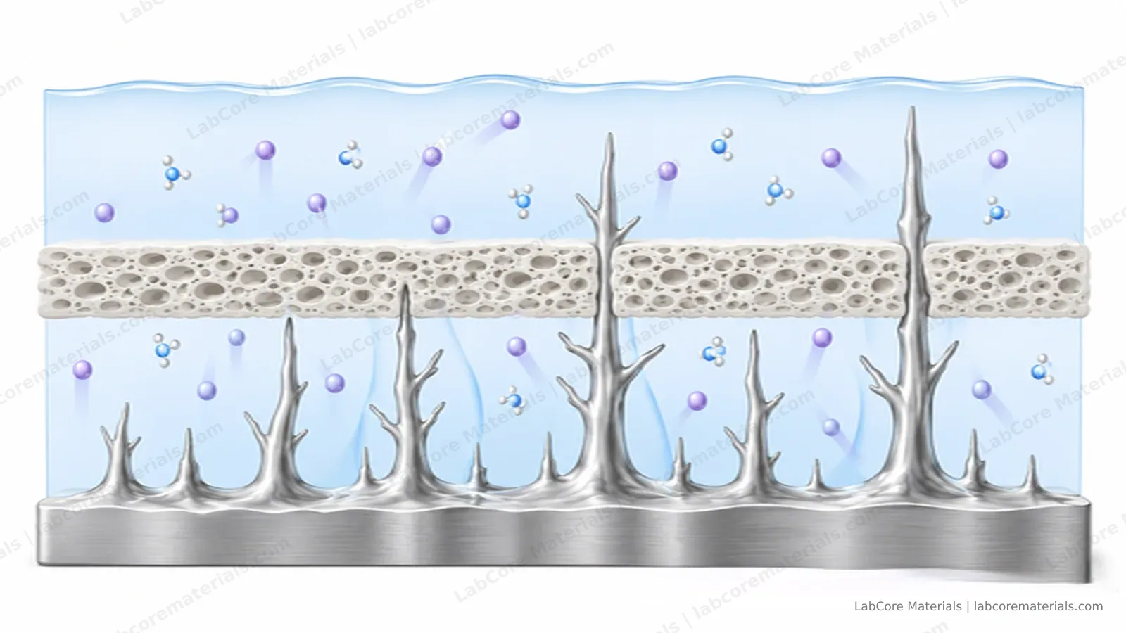
Dendrites grow along the separator's pores. Before penetration there are usually warnings: rising self-discharge, abnormal end-of-charge voltage behavior, sliding Coulombic efficiency — signs of a soft short formed by partial dendrite contact. A hard short follows when the electrodes touch directly: large currents through a local hot spot, and the window for thermal failure opens from that moment. Ceramic-coated separators raise the threshold and buy time; they do not remove the endpoint.
Seeing it before the short
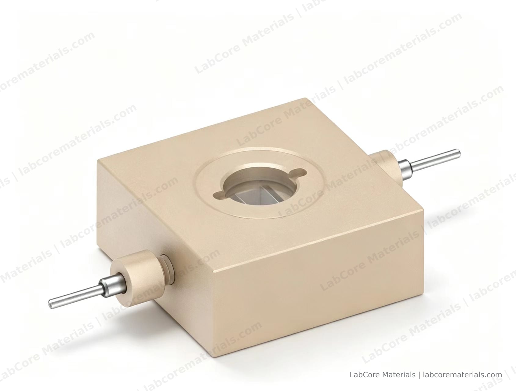
Indirect indicators all lag: by the time Coulombic efficiency drops, self-discharge rises or EIS spectra deform, the dendrite is already established. To intervene at the nucleation stage, the most direct approach is to watch the anode surface. Optical in-situ observation distinguishes uniform deposition from mossy deposits and true dendrites — and morphology is exactly what you need to grade the risk. Before a dendrite becomes a failure, optical methods are among the few that can catch it early; the in-situ lithium dendrite observation cell provides a sapphire window for the purpose: cycling and microscopy run simultaneously, without interrupting the experiment or opening the cell.
To pair the morphology with the potential response of both electrodes, the visible three-electrode Swagelok-type test cell puts an optical window and a reference electrode in the same body — plating diagnosis is complete only when morphology and potential agree.
Applications
| Source | Setup | Observation |
|---|---|---|
| EL-CELL GmbH. Lithium metal dendrites: pictures speak louder than words [Application note]. 2017. el-cell.com | Sapphire-window optical cell with lithium metal as reference electrode | Dendrite growth and shrinkage recorded in real time over cycling; most dendrites remained after stripping, and growth to the counter electrode ended in an internal short |
| Choi T Y, Kim J, Muralidharan V, et al. Depth-to-Areal Staging Heterogeneity Preceding Lithium Plating in Graphite Anodes Revealed by Operando Imaging. Advanced Science, 2026. DOI: 10.1002/advs.77918 | Operando optical cell with a lithium reference electrode; graphite anode | Quantitative imaging of lithiation depth and in-plane distribution before and after plating onset, with morphology and electrochemical data acquired simultaneously |
Conclusion
In-situ observation turns dendrite formation from something inferred after failure into something recorded while it happens. By following nucleation sites, morphological evolution and detachment continuously, with the electrode potential acquired in parallel, the interfacial kinetics of plating and stripping become analysable: why nucleation favours particular sites, how morphology shifts between diffusion-controlled and reaction-controlled growth, and how dead lithium is generated and accumulates during stripping. The resulting correspondence between process conditions, interfacial behaviour and failure path shows which link in the mechanism a given electrolyte, coating or pressure scheme actually changes — rather than only how many cycles it buys.
For lithium-metal anodes, fast-charging systems and solid-electrolyte interface research, such data provide evidence at the mechanistic level: which failure paths are suppressed, and which are merely postponed. The observation window therefore belongs inside the cycling test rather than after it.